
Glaucoma: The Silent Thief of Sight — Why Early Detection in Durban Saves Vision
23 June 2026
Macular Degeneration: Understanding the Leading Cause of Vision Loss After 50
23 June 2026

Diabetes and Your Eyes: Why Every Diabetic in KZN Needs an Annual Eye Exam
Diabetes does not just affect your blood sugar. It is quietly, persistently damaging the blood vessels at the back of your eyes — and in most cases, you will not feel a thing until serious harm has already been done.
Diabetic retinopathy is the leading cause of new blindness in working-age adults worldwide. One in three people living with diabetes will develop some form of it. And yet the condition is almost entirely preventable with one simple intervention: an annual dilated eye examination.
If you have Type 1 or Type 2 diabetes, this article explains what diabetic eye disease is, why it matters, and what an ophthalmologist looks for during the exam that could save your sight.
How Diabetes Damages the Eyes
Chronically elevated blood sugar damages the small blood vessels throughout the body. In the eye, the retina — the delicate neural tissue that lines the back of the eye and processes everything you see — is especially vulnerable. When the tiny retinal blood vessels weaken, they can leak fluid, bleed, or close off entirely. In advanced stages, the eye responds by growing abnormal new blood vessels that are fragile and prone to sudden, severe bleeding.
This process is called diabetic retinopathy. It progresses through distinct stages, from mild (small microaneurysms), through moderate and severe (increasing haemorrhages and vessel damage), to proliferative diabetic retinopathy — the most dangerous stage, where abnormal new vessels can bleed into the eye or cause the retina to detach.
The Danger of Diabetic Macular Oedema
At any stage of diabetic retinopathy, fluid can leak into the macula — the part of the retina responsible for your sharp, central vision. This condition, known as diabetic macular oedema (DMO), is the most common cause of vision loss in people with diabetes. It causes blurring and distortion of the central vision you rely on for reading, driving, and recognising faces.
The critical point is this: DMO can develop even in early-stage retinopathy, when you would have no reason to suspect anything is wrong. Only a dilated eye examination can detect it.
Why You Cannot Rely on Symptoms
In its early and moderate stages, diabetic retinopathy typically produces no symptoms at all. Your vision may feel perfectly normal while significant damage is accumulating in the retinal blood vessels. By the time symptoms appear — blurred vision, dark spots, or sudden vision loss from a bleed — the disease has often reached an advanced stage where treatment is more complex and outcomes less certain.
What Happens During a Diabetic Eye Exam?
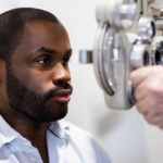
A comprehensive diabetic eye examination with a specialist ophthalmologist typically includes:
- A visual acuity test to assess your current level of vision
- Pupil dilation using eye drops so the ophthalmologist can see the full retina clearly
- A dilated fundus examination — a direct, detailed inspection of the retina, optic nerve, and blood vessels
- Optical coherence tomography (OCT) — a non-invasive scan that measures retinal thickness with extraordinary precision, detecting macular oedema even before it affects your vision
- Fundus photography to document the current state of your retina for future comparison
In some cases, fluorescein angiography may be used. This involves injecting a harmless dye into the arm and photographing how it moves through the retinal blood vessels, revealing any areas of leakage or abnormal vessel growth.
The entire examination takes approximately 30 to 60 minutes. The dilation drops will blur your near vision for a few hours, so it is advisable to arrange transport home.
How Diabetic Eye Disease Is Treated
The single most powerful treatment for diabetic eye disease is not a procedure — it is systemic control. Keeping your HbA1c below 7%, maintaining healthy blood pressure, and managing cholesterol levels dramatically reduce the risk of retinopathy developing or worsening.
When retinopathy does require ophthalmic treatment, the options are highly effective. Anti-VEGF injections are the standard of care for diabetic macular oedema and proliferative retinopathy. These injections, administered directly into the eye under local anaesthetic, block the growth factor responsible for abnormal blood vessel development and leakage. Laser photocoagulation — either targeted macular laser or panretinal laser — remains an important tool, particularly in advanced disease. In severe cases involving vitreous haemorrhage or tractional retinal detachment, vitrectomy surgery may be necessary.
The earlier diabetic eye disease is detected, the simpler and more effective the treatment. This is the fundamental case for annual screening.
The South African Context
Approximately 12% of South African adults live with diabetes — and that number is growing. In KwaZulu-Natal, the combination of high diabetes prevalence and limited access to specialist eye care in many areas means that too many patients present with advanced disease that could have been prevented.

Dr Johann Snyman is a specialist ophthalmologist in Kloof and Westville, Durban, with a particular commitment to diabetic eye care. He provides comprehensive diabetic eye screening and treatment, including OCT imaging, laser therapy, and anti-VEGF injection therapy. His practice works closely with general practitioners, endocrinologists, and diabetologists across KZN to ensure patients receive timely, coordinated care.
As the founder of Africa20twenty — a non-profit providing free cataract surgery in underserved rural communities — Dr Snyman understands that access to specialist care should not depend on geography or income.

If You Have Diabetes, Your Eyes Need Annual Screening
Don’t wait for symptoms. A single appointment can detect changes that, caught early, are almost always treatable.
Book an appointment · +27 (0)31 854 0003