
Red Eye: When It Is More Than Conjunctivitis
29 October 2026
High Blood Pressure and Your Eyes: What the Retina Can Reveal
29 October 2026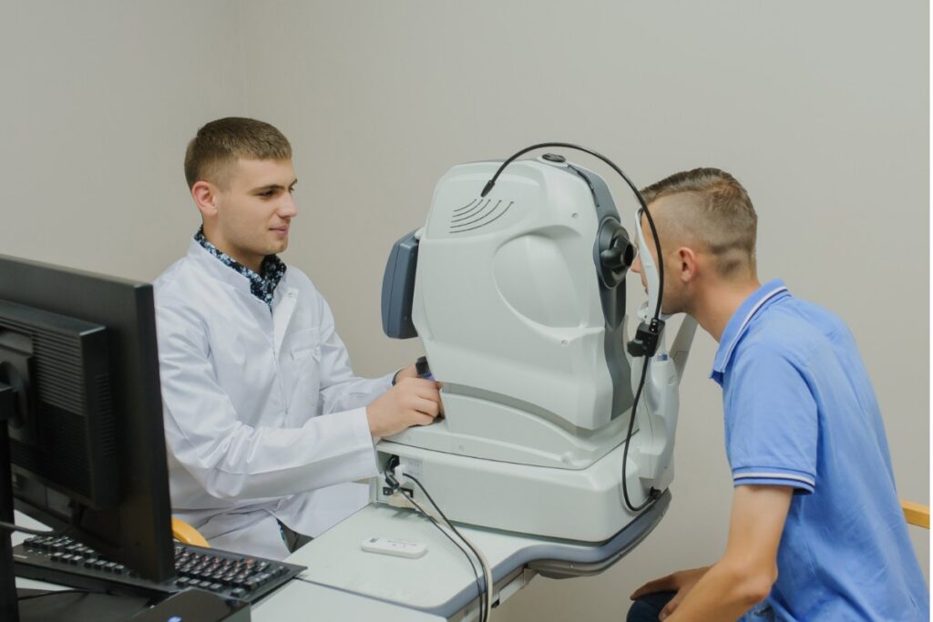
OCTOBER 2026 · ARTICLE 03 OF 04 · UNDERSTAND
What Is an OCT Scan? Seeing What a Vision Chart Cannot
DR JOHANN SNYMAN · OPHTHALMOLOGIST · KLOOF & WESTVILLE · 4 MIN READ
A vision chart tells us how small a letter you can see. It does not tell us everything about why you can (or cannot) see it.
Modern ophthalmology therefore relies increasingly on imaging that can examine the microscopic structures responsible for vision. One of the most useful of these tests is optical coherence tomography, usually shortened to OCT.
An OCT scan uses light waves to build highly detailed cross-sectional images of structures inside the eye. It is non-invasive, quick and does not use ionising radiation.
An OCT does not simply photograph the eye. It allows us to examine its layers.
Looking Beneath the Surface of the Retina
The retina is not a single sheet. It consists of multiple specialised layers working together to convert light into the electrical signals that eventually become vision.
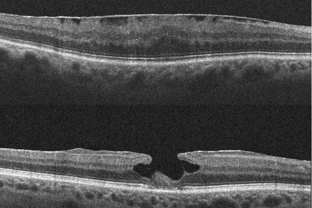
An ordinary retinal photograph shows the surface appearance of the retina. OCT adds a different perspective, creating slices through the tissue so that an ophthalmologist can see changes in thickness and architecture.
This is especially valuable at the macula, the small central area responsible for reading, recognising faces and seeing fine detail.
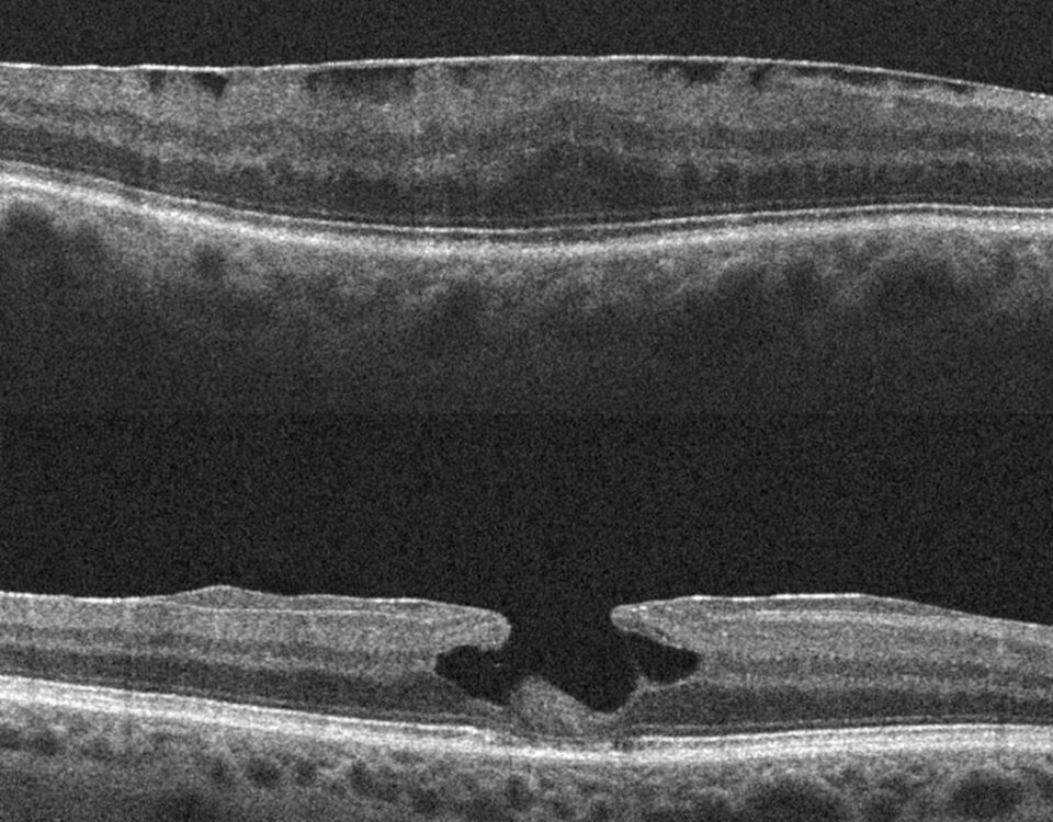
Fluid from diabetic macular disease, changes related to macular degeneration, a macular hole, traction on the retina or a thin epiretinal membrane may all be demonstrated in far greater detail on OCT than they can be from a vision chart alone.
OCT and the Optic Nerve
OCT is equally important in conditions affecting the optic nerve.
In glaucoma, for example, damage may gradually reduce the thickness of the retinal nerve fibre layer and ganglion cell complex. Measuring these structures allows the ophthalmologist to compare your results with previous scans and look for meaningful change over time.
This is why one scan is useful, but a series of accurately repeated scans can be even more informative.
A result that is stable over several visits tells a different story from one showing progressive thinning.
Why Good Vision Does Not Always Mean a Normal Scan
Some eye conditions alter anatomy before the patient notices a major change in everyday vision.
A person may still read a relatively good line on the chart despite early structural changes. Conversely, an OCT abnormality does not automatically mean that vision is about to deteriorate.
The image has to be interpreted together with your symptoms, clinical examination, eye pressure, visual field testing and other findings.
Technology supplies information. The diagnosis still depends on clinical judgement.
What Happens During an OCT Scan?
You sit in front of the OCT machine and look at a target while the instrument scans the eye. Nothing touches the eye during a standard retinal OCT.
The scan itself takes only moments, although several images may be repeated to obtain the most reliable result. Pupil dilation is not always required for OCT, but it may be used as part of the wider examination.
Very dense cataracts, poor fixation or other optical problems can reduce image quality. Automated measurements can also occasionally misidentify retinal boundaries, which is another reason the scan must be interpreted rather than simply accepted as a number on a screen.
Why Baseline Imaging Matters
A baseline scan gives your ophthalmologist something invaluable: a personalised reference point.
If you return months or years later, subtle changes can be compared with your own earlier eye rather than relying only on a population average.
For diseases that progress slowly, this ability to measure change can influence decisions about when to observe, investigate further or adjust treatment.
See More Than the Surface.
Modern imaging does not replace careful examination. It extends it, helping turn tiny structural changes into information that can be measured, compared and understood.
Medical information cross-checked against current ophthalmology references describing OCT use in retinal, macular and optic nerve disease.
Related reading
If you have concerns about your eyes, Dr Johann Snyman’s Kloof and Westville rooms are here to help.